From the moment the idea arose to tackle the issue originating from Erasmus MC, it…
The struggle of experiments
The struggle of experiments
It seemed unmistakably simple: just set up several nebulizer systems and investigate the airflow of the aerosol. This way it would be easy to find out what nebulizing system exposes its environment as little as possible. How hard can it be? As it turns out, setting up an experiment is never straightforward.
From this initial idea many questions dashed through the brainstorms of the dedicated team members. How can we visualize aerosol and flow through the system? Will this airflow be affected by room ventilation or draughts? Should the breathing of a patient be simulated? How are measurements going to be compared? – But this is just the tip of the iceberg.
Over half of the summer we dug through most of the iceberg, currently finishing up the procedure for carrying out experiments. It took several iterations of research proposals, scrapped ideas and lots of meetings with experts across the whole country. Their expertises range from cardiovascular biomechanics to building physics! At first sight, these areas might not seem relevant for our research. However, the opposite is true.
Meet Veronica
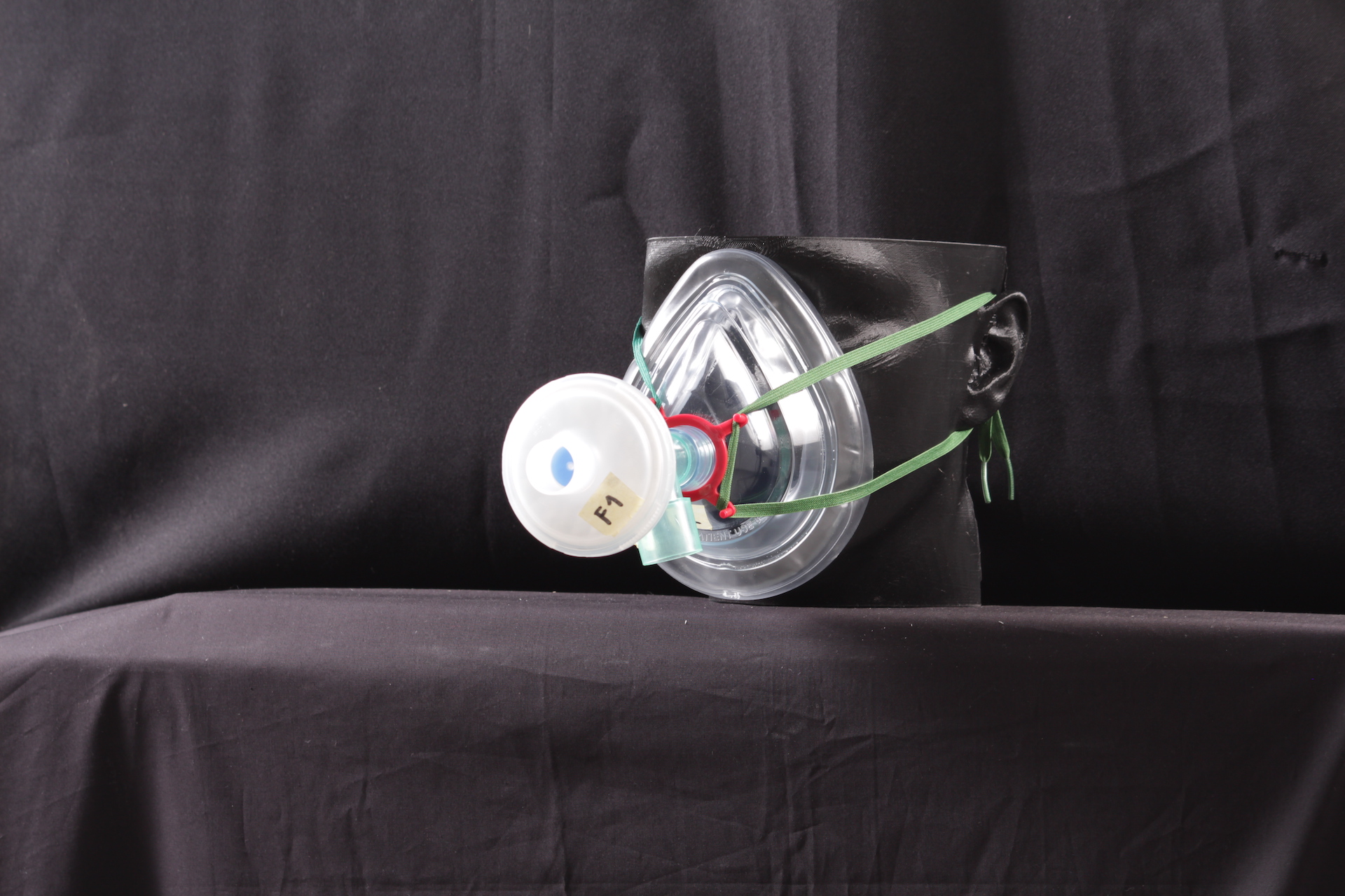
For example, Marcel Rutten (part of the TU/e research group Cardiovascular Biomechanics) provided a specialized pump which is normally used to simulate a heart which was modified to simulate the breathing of a patient receiving treatment. By attaching this to a 3D printed head we created it: the simulated patient that we with respect refer to as Veronica. She is the brave test subject who is going to be exposed to several experiments, and the first part of our setup.
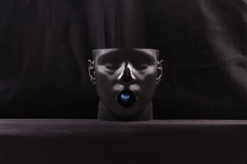
Veronica needs a place to live. Luckily, we found the perfect place for her, someplace where it is never too cold nor too hot. The building physics and science laboratory in the Vertigo building at the TU/e Campus has a chamber which has full control over the temperature and ventilation (for a reason!). The head of this laboratory, Jan Diepens, agreed to house Veronica during the summer for these experiments. We belief she quite likes the place! More on this can be read in this previous blog post. Since the simulated patient and the lab is arranged, only several different matters need to be taken care of…
Looking for aerosol
Well, medical aerosols are, sadly, not clearly visible to the naked eye. So, analyzating the flow of aerosol through a nebulizing system would be nearly impossible. Therefore, we switched out the aerosol generator with an industrial smoke generator. This device creates similarly behaving aerosol in far higher quantities to increase the visibility.
But visibility is not the only requirement, since ‘eye-balling’ is not a part of the scientific method. Just as described in literature, the smoke is used such that we can analyze images taken during experiments. This required some alterations to Veronica’s living area. With help of Dekate Mousa, the student film and photography association of Eindhoven, it has become a professional photo studio.
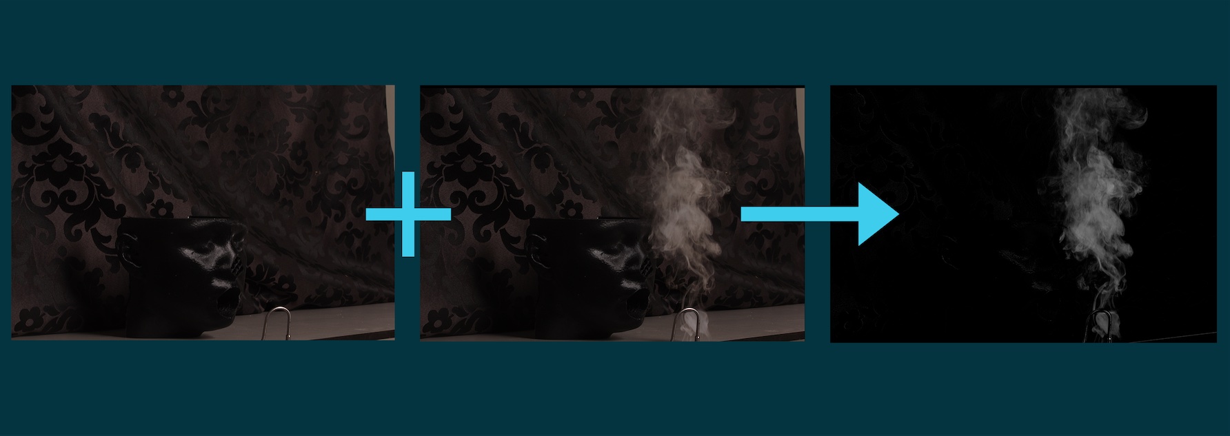
Even though Veronica would make an amazing photomodel, she will not be visible in the photos. During the analysis of these experimental results, we will be looking for the changes in an image with respect to a reference image. So, only if smoke is added in the frame of a photo this will be visual in the final analyzed photo! From this analyzed photo we can metric the results. All in all, you can imagine that it is a long way from stepping into the lab to getting results.
The experiment
We start off the day with a smile and meeting up in the lab in Vertigo. The nebulizing system is prepared just as all the equipment in the lab. This is probably also the time for some chit-chat with the lovely lab employees, which are the greatest help if we for example need help with the equipment. First, the photo–equipment gets turned on. Secondly, the breathing simulator is initialized. Veronica gets equipped with the nebulizes system. Then, at last, the smoke generator is attached to the mask if everything and everyone is ready to start the experiment.
The smoke starts flowing down a tube into the nebulizing system. The simulated breathing of Veronica makes some of the smoke gush though opening between the nebulizer mask and her face. That is captured by periodically captured photos. The photos are showing jets of smoke. After some time, the room fills up with smoke and the measurement ends. This is where the ventilation control of the lab comes into play, refreshing the air in the room in merely 15 minutes. When all smoke has found a way out of the room a last photo is taken as reference. Now the data analysis can start. Using powerful photo analysis tools, we can ‘subtract’ the reference photo from the photos which show jets of smoke. This leaves an image which only shows smoke.
In this way we can compare the aerosol released for different nebulizer systems. That is the focus of this experiment. Finding out which nebulizing system exposes its environment the least to aerosol, which can possibly contain unwanted particles like the SARS-COV-19 virus.
That is our first step in trying to make aerosol treatment safer. The struggles we experienced went even further than described here, the iceberg was much bigger than we thought. There were problems with visualizing the smoke, printing the 3D-models, connecting all tubing, technical problems and much more to name.
Soon the first experiments will be conducted. We are sure to run into more challenges, but as we have gone through this struggle before: challenges like these are inherent to the scientific method but luckily also where we learn the most!
When all these experiments are done, the next step is to investigate the aerosols generated by different nebulizer systems. When the time comes, a new blog will be posted. Stay in the loop by following our LinkedIn.
Gerelateerde berichten